Adam Sewell, M.D.
RESOURCES
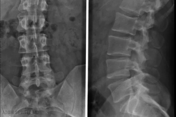
DISCOGRAPHY
Discography is a diagnostic tool used to determine whether certain discs of the spine are the source of a patient’s neck or back pain. Most of the population experiences low back pain, at least, sometime during their lifetime. It is the fifth leading cause of medical visits and the leading cause of workplace-related disability. Discography is used to pinpoint the cause of low back and neck pain, especially when non-invasive imaging, such as magnetic resonance imaging (MRI), has failed to reveal the source of pain (Wichman 2007). Discography is considered for patients with idiopathic (uknown), but disabling, neck and back pain.
Understanding the anatomy and function of the spine is important in evaluating back pain. The spine is made up of vertebrae (bones of the spine) that provide a flexible support for the back muscles and spinal cord. Certain conditions cause pain in, and around, the spine, including Spinal stenosis, vertebral body fractures, Osteoporosis, Osteoarthritis, Spondylolisthesis (scar tissue), neoplasms (Primary vs. metastatic lesions), and infections. Separating each individual vertebrae are discs that act as cushions to minimize the impact that the spinal column receives. The softness of the discs give them a tendency to herniate posterior (backwards) through the outer disc segment and ligaments. Bulging and leaking discs often cause irritation to the adjacent nerves. This results in disc disease, which accounts for about 10% of all doctor visits for low back pain. It can be acute, resulting from herniation or trauma, and can become chronic. Degenerative disc disease is progressive, causing a thinning and degeneration of the discs over time.
Discography Procedure
After you skin is thoroughly cleaned, s small needle will numb the area with a local anesthetic. A larger needle will then be inserted and positioned near the outer layer of the disc. Fluoroscopy, real time X-ray, is used in order to assure proper placement of the needle. A contrast solution is injected into the disk and your response to the injection is observed at different locations. If pain is experienced, then it is possible that your doctor has located the source of your pain. This process is typically repeated twice for maximum relief. The procedure usually takes less than an hour. A CT scan may be ordered directly after the procedure to ensure that the contrast die has spread properly.
Discography Benefits
Although MRI is considered a very good method for showing disc abnormalities, it does not show direct causes of pain. A study in 2007 revealed that Discography is more accurate at locating disc pain and ruptures than is MRI. This is true for similar conditions as well (Montes Garcia 2007). Since discography is considered a minimally invasive procedure it is performed when your physician highly suspects the cause of your pain. Generally, when students undergo discography, they have tried a myriad of other ineffective treatments for isolating the precise area of their pain. An MRI and CT scan can show abnormalities, but a discography reveals the exact location of the pain. Sometimes, moderately effected discs can cause severe pain, and run the risk of being treated improperly if based solely on MRI and CT findings (MRI/CT scan).
Discography Risks
As with all treatments, there is risk. However, the risk with discography is usually small. Although very uncommon, discitis (infection of the disc) can be severe. Other risks include bleeding, hematoma, headache, and increased pain.
Discography Outcome
Once your discography has been performed and your doctor has been able to isolate the source and location of your pain, you will be described an effective treatment plan. Discography is believed to be the best source of investigative protocol for disc disease (Buenaventura 2007). If you are suffering from chronic back pain that has been refractory to other treatments contact Arkansas Pain Specialists today to see if you can benefit from discography, or any of their other innovative, pain-relieving services.
Journal Articles
Why Choose Adam Sewell, M.D.?
we believe in treating the whole person, not just the symptoms. Our approach to medicine is integrative, combining the best of modern science with personalized care to deliver real, lasting results. We work closely with each patient to develop a customized treatment plan that aligns with their health goals and lifestyle.
Patient-Centered Care
We listen to your concerns, understand your goals, and work with you to create a treatment plan that's right for you.
Advanced Technology
We utilize the latest medical technologies and techniques to provide you with the most effective treatments available.
Expertise You Can Trust
Dr. Adam Sewell is a triple board-certified physician with a passion for helping patients achieve their best health. His expertise in regenerative medicine, hormone therapy, and performance medicine ensures that you're in capable hands.
Comprehensive Health Solutions
From gut health to cognitive function, joint regeneration to hormone optimization, our services are designed to address every aspect of your health.

Ready to Take the Next Step?
If you're ready to transform your health and live your best life, we're here to help. Contact us today to schedule a consultation and discover how Adam Sewell, M.D. can help you achieve your health goals.
Contact Us
Please feel free to reach out to us at adam@adamsewellmd.com or kindly complete the form. Rest assured, we will respond to your inquiry at the earliest opportunity.
Our Locations

Stay in the Know with Our Newsletter!

Copyright © 2025 Adam Sewell M.D., All rights reserved.